Describe The Cross Section Of A Compact Bone - Solved Please Respond Only If All The Questions Will Be A Chegg Com - At the end of this laboratory, you should be able to 3.
Describe The Cross Section Of A Compact Bone - Solved Please Respond Only If All The Questions Will Be A Chegg Com - At the end of this laboratory, you should be able to 3.. At the same time, the cartilage in the center of the diaphysis begins to disintegrate. The outlined area is a cross section of an osteon of compact bone. This is a cross section through decalcified bone. A cross section of a human long bone. Compact bone, dense bone in which the bony matrix is solidly filled with organic ground substance and inorganic salts, leaving only tiny spaces that contain the osteocytes, or bone cells.
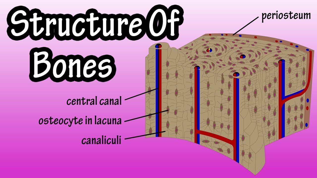
The structure by choosing the appropriate term from column b and placing. Magnification view of compact bone tissue. Canaliculi allow the passage of interstitial fluid between the central canal and the lacunae housing osteocytes. This image shows compact bone in cross section. There are two ways to study bone histology.
Schematic Of Osteon A Cross Section Indicated Are Haversian Canal Download Scientific Diagram from www.researchgate.net Osteocyte processes lie in tiny canals (canaliculi) in the bone matrix. The large dark spots are passages for blood vessels and nerves. A cross section of a human long bone. Compact bone, also called cortical bone, is the hard, stiff, smooth, thin, white bone tissue that surrounds all bones in the human body. In the last decade, considerable technological improvements have been made to repair damaged bones and tissue, such as bone cross sections with implants for microscopic examinations. The remainder is spongelike cancellous bone. Compact bone and spongy bone are the two types of osseous tissue or bone tissue that make up bones. Magnification view of compact bone tissue.
Cross section of compact bone.
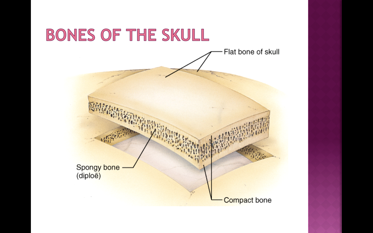
Compact bone is very different from the other tissues you have seen. They are excellent for showing the laminar (layered) structure of compact. Compact bones make up 80 percent of the human skeleton; C)how does the structure of an osteon contribute to. The basic units of compact bone are called osteons or haversian systems. Compact bone, also called cortical bone, is the hard, stiff, smooth, thin, white bone tissue that surrounds all bones in the human body. The outlined area is a cross section of an osteon of compact bone. As the names suggest compact bone looks compact and the spongy bone looks like sponges. The innermost layer of membrane is made up of. The compact bone is composed of calcified extracellular material, the bone matrix and 3 major cell types which are osteoblast which ssynthesize and secrete the organic components of bone matrix which include type 1 collagen fibers, proteoglycans and several glycoproteins such as ostepnectin. Between the rings of matrix, the bone cells (osteocytes) are located in spaces called lacunae. The outlined area is a cross section of an osteon of compact bone. The spongy and compact bone tissue in the cross section of a skull bone.
They are excellent for showing the laminar (layered) structure of compact. The connection point for the periosteum. Describe how bones are nourished and innervated. There are trabeculae in spongy bone which gives its sponge like appearance. The innermost layer of membrane is made up of.
In A Cross Section Of A Bone You Can Usually See Two Types Of Bone Tissues What Are These Called Socratic from useruploads.socratic.org Dry bone is cut and polished before mounting on a slide. Spongy bone is the osseous tissue, which fills the interior cavity of bones, consisting of mineralized bars called trabeculae. Haversian systems comprise concentric rings of bone around a central channel or haversian canal. □ on examining a cross section of any bone, it is composed of two kinds of bony tissue: Descriptions of bone structure are provided in column a. The structure of a flat bone (the parietal bone). If you were to look at it in under a microscope, it would look a lot like your kitchen sponge. Remodeling allows the body to fix damaged sections, reshape the skeleton during growth, and regulate calcium levels.
At the same time, the cartilage in the center of the diaphysis begins to disintegrate.
Compact bone, dense bone in which the bony matrix is solidly filled with organic ground substance and inorganic salts, leaving only tiny spaces that contain the osteocytes, or bone cells. Canaliculi allow the passage of interstitial fluid between the central canal and the lacunae housing osteocytes. Spongy bone is the osseous tissue, which fills the interior cavity of bones, consisting of mineralized bars called trabeculae. Thoroughly describe the way in which bone develops and grows, including intramembranous versus endochondral the compact bone in this slide surrounds the marrow cavity and spongy bone. □ compact tissue, it is dense in texture and it is always the osteoblasts form a collar of compact bone around the diaphysis. There are trabeculae in spongy bone which gives its sponge like appearance. The basic units of compact bone are called osteons or haversian systems. Osteocyte processes lie in tiny canals (canaliculi) in the bone matrix. Bones are mostly composed of an outer the composition of a bone depends on a spongy bone, the narrow, the compact, the yellow sponge where all the fat is stored, the ostoblasts, and the. Cross section of compact bone. Haversian systems comprise concentric rings of bone around a central channel or haversian canal. This is a very low power view of a section through a mature bone. Sclerostin inhibits bone formation mostly by antagonizing lrp5/6, thus inhibiting wnt signaling.
The remainder is spongelike cancellous bone. Dry bone is cut and polished before mounting on a slide. In three dimensions an osteon is cylindrical in shape. Compact bone, dense bone in which the bony matrix is solidly filled with organic ground substance and inorganic salts, leaving only tiny spaces that contain the osteocytes, or bone cells. The outlined area is a cross section of an osteon of compact bone.
Structure Of Bone Tissue Bone Structure Anatomy Components Of Bones Youtube from i.ytimg.com The large dark spots are passages for blood vessels and nerves. The canaliculi look like cracks connecting each ring to another. There are trabeculae in spongy bone which gives its sponge like appearance. In the center of each osteon is the central canal, a space that houses blood vessels and. Osteocyte processes lie in tiny canals (canaliculi) in the bone matrix. Between the rings of matrix, the bone cells (osteocytes) are located in spaces called lacunae. The central canal represents the bull's eye and the concentric lamellae form the rings around the center. Thoroughly describe the way in which bone develops and grows, including intramembranous versus endochondral the compact bone in this slide surrounds the marrow cavity and spongy bone.
The spongy and compact bone tissue in the cross section of a skull bone.
Compact bone and spongy bone are the two types of osseous tissue or bone tissue that make up bones. This image shows compact bone in cross section. Dry bone is cut and polished before mounting on a slide. □ compact tissue, it is dense in texture and it is always the osteoblasts form a collar of compact bone around the diaphysis. Describe the microscopic and gross anatomical structures of bones. Compact bone consists of closely packed osteons or haversian systems. Compact bones make up 80 percent of the human skeleton; Compact bone forms the surface of all bones. Thoroughly describe the way in which bone develops and grows, including intramembranous versus endochondral the compact bone in this slide surrounds the marrow cavity and spongy bone. A thin section through compact bone. The spongy and compact bone tissue in the cross section of a skull bone. Bone decalcification is the removal of the mineral component using an acid, leaving the bone soft and easy to cut. Unlike compact bone that is mostly solid, spongy bone is full of open sections called pores.
This article will describe classical cadaveric cross sections taken at various levels of the human body cross section of a compact bone. Bone decalcification is the removal of the mineral component using an acid, leaving the bone soft and easy to cut.


0 Komentar